
Forensic Science / Crime Labs
Reports, presentations, and other files may be included in these PAX-it case folders, for total project management.
PAX-it’s network site license allows shared–or separate–databases for Trace Evidence, Firearms & Toolmarks, Documents, Drug Analysis, DNA lab, Serology, Coroner’s/Medical Examiner’s office, and other departmental needs.
- Image annotation tools, as overlays, to point out features in reports or presentations
- Image blending tools for comparisons and alignment of markers
- Overlays of images, grids, markers, etc on images
- Side-by-side construction of plates for reports or presentations
- Prefill of database information per case, as images are brought in
- Auto-sorting of images into case folders by case number, or other designated sort function
- Easy report preparation: With one click, Create and customize report layouts, and generate those reports in Microsoft Word or Excel
- Easy presentations and sharing: With one click, export images –with or without annotation layers– to PowerPoint or to email
- Network Site Licensing for powerful, immediate sharing of assets
Worried about the security of your digital assets? The PAX-it Extended Security Module integrates additional layers of protection for you digital assets. Features include:
- Unique logins and administrative tools
- Electronic records and electronic signatures
- Detailed audit trails
- PAXcam cameras for high resolution imaging from microscopes and macrostands
- Calibrated measurements from forensic images: linear, area, angle, arc, and more
- Image analysis software for auto detection of objects, counting, sizing, area fractions, etc. Easy to use reporting function includes report generation in MS-Excel for application of formulas, graphing of data, conditional formatting, and more
- QuickSolve conversion utility to bring legacy databases into current PAX-it format
Forensic Science Images
Images taken with PAXcam cameras from various labs and samples.
NOTE: Images have been resized downward, and compressed, in order to present them as samples on the web. Original images are larger files and higher resolution.
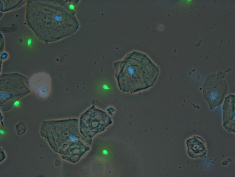
PAXcam2+, Phase contrast and fluorescence images are blended in PAX-it to show the location of the fluorescing sperm heads
PAXcam2+, Phase contrast and fluorescence images are blended in PAX-it to show the location of the fluorescing sperm heads
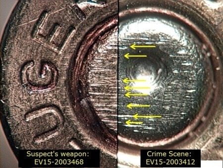
a photo taken with a Paxcam, of the breechface match from comparison scope
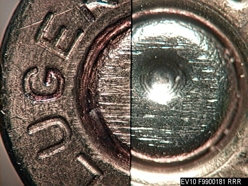
a photo taken with a Paxcam, of the breechface match from comparison scope
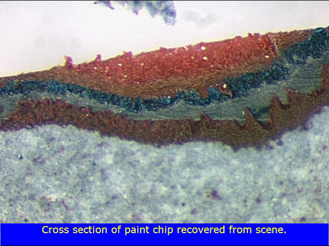
Cross section of paint surface, reflected light compound microscope, annotated in PAX-it
Cross section of paint surface, reflected light compound microscope, annotated in PAX-it
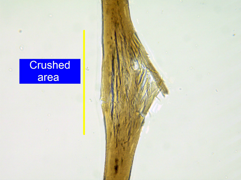
PAXcam2; Crushed hair, reflected light compound microscope, annotated in PAX-it
PAXcam2; Crushed hair, reflected light compound microscope, annotated in PAX-it
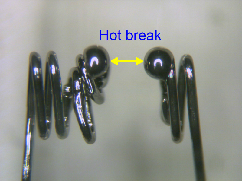
PAXcam2; Headlight filament break, stereozoom microscope, annotated in PAX-it
PAXcam2; Headlight filament break, stereozoom microscope, annotated in PAX-it
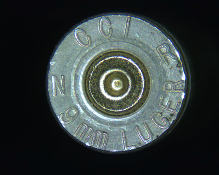
PAXcam3; Luger

PAXcam ARC; Winchester
PAXcam ARC; Winchester